Unlock fMRI versus MRI: A Clinician's Guide to Imaging Choices
Mar 15, 2026
When you’re weighing fMRI versus MRI, the choice boils down to a fundamental question: are you looking at the brain’s structure or its function? A standard MRI scan gives you a static, high-resolution anatomical blueprint. It’s your go-to for spotting physical issues like tumours, injuries, or lesions. An fMRI, on the other hand, creates a dynamic activity map, showing you which parts of the brain are lighting up and working in real time.
FMRI Versus MRI: Answering the Core Clinical Question
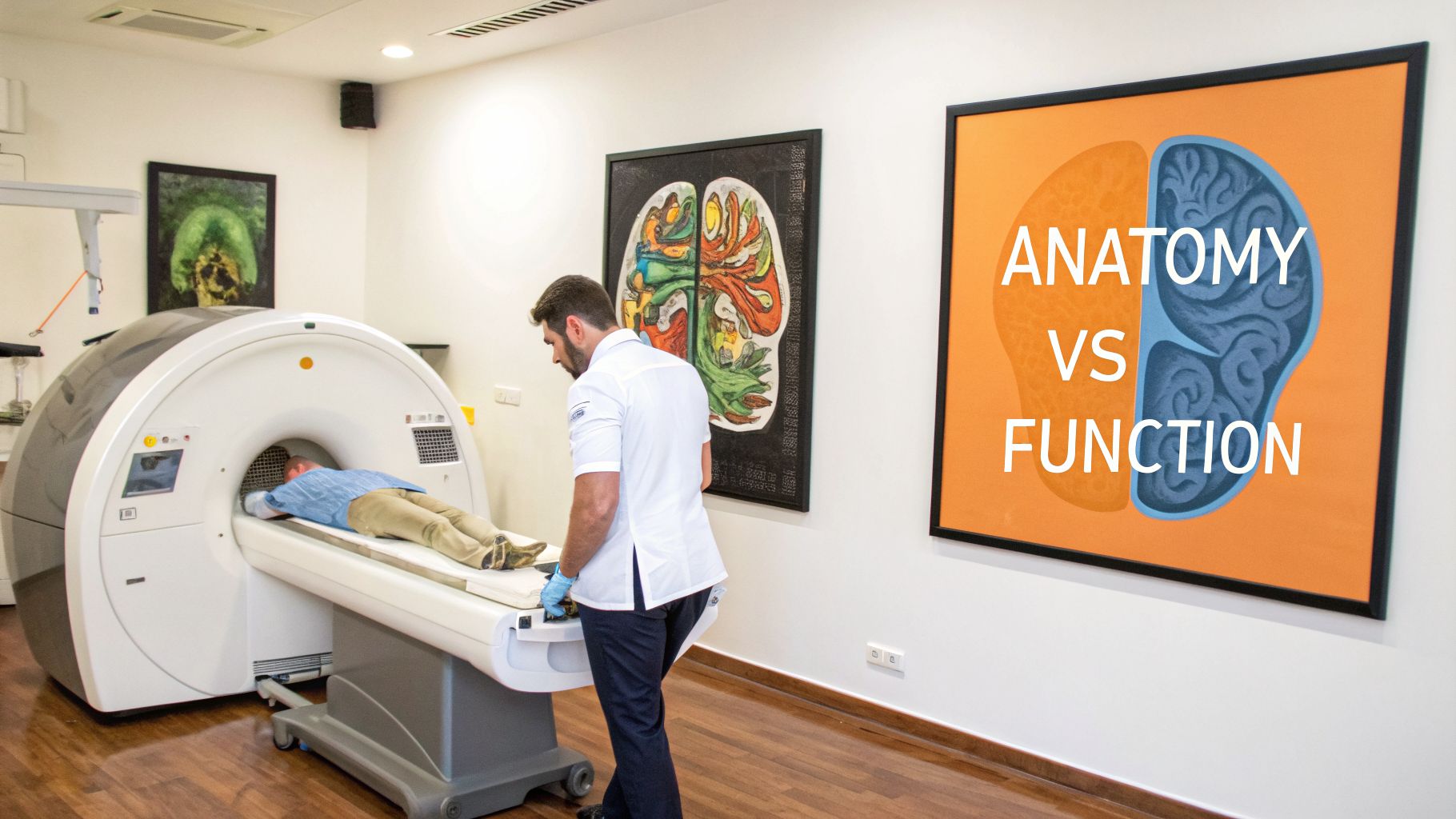
Think of it this way: an MRI gives you a beautifully detailed street map of a city, while an fMRI shows you the live traffic patterns—where the cars are moving, where the jams are, and which routes are most active. This core difference is what guides every clinical decision about which scan to order. For example, if a patient presents with sudden weakness on one side of their body, your immediate question is structural: "Is there a bleed or a blockage?" An MRI is the actionable choice to get that answer.
So, if your clinical question is about the brain’s physical state, an MRI is your primary tool. It's the definitive method for confirming or ruling out a structural abnormality you might suspect based on a patient’s symptoms.
A great way to frame it is that an MRI reveals the 'what' and 'where' of brain anatomy. In contrast, an fMRI starts to uncover the 'how' of brain function. An MRI can confirm a lesion is present; an fMRI can show how the brain is working around that lesion.
This distinction is what helps us move from a general concern to a targeted diagnostic plan. While both technologies rely on the same powerful magnetic hardware, they’re calibrated to answer entirely different questions about a patient's brain health.
Quick Comparison: MRI vs fMRI at a Glance
For a quick, scannable reference, the table below breaks down the essential differences. It's a handy tool for quickly aligning your clinical question with the right imaging technology.
Attribute | Structural MRI (The Brain's Blueprint) | Functional MRI (The Brain's Activity Map) |
|---|---|---|
What It Measures | The static physical structure of brain tissue (anatomy). | Changes in blood-oxygen levels as a proxy for brain activity. |
Primary Purpose | To diagnose structural issues like tumours, strokes, or injuries. | To map brain function for research or pre-surgical planning. |
Data Output | High-resolution, static 3D images of the brain. | Dynamic, lower-resolution maps showing brain activity over time. |
While this table simplifies the concepts, the rest of this guide will dig into the nuanced scenarios where these differences truly matter. Understanding these initial distinctions is the first step, but diving deeper into specific conditions can provide even more context. For instance, exploring the role of a brain scan for ADHD can clarify when functional imaging might fit into a broader assessment.
Understanding the Science Behind Each Imaging Technology
To really get to the heart of what separates an MRI from an fMRI, we have to look at how they each see the brain. Think of it like this: one is a high-resolution photograph, and the other is a live-action video. That’s the core difference, and it all starts with the underlying physics.
A structural MRI works like a detailed anatomical mapmaker. It uses powerful magnetic fields to line up the hydrogen atoms found in the water of your brain’s soft tissue. When the machine sends out a pulse of radio waves, these atoms get knocked out of alignment for a moment. As they snap back into place, they emit signals that the MRI scanner translates into a stunningly detailed, static 3D image.
This is why MRI is the gold standard for visualizing brain anatomy. It gives clinicians a crystal-clear, static "blueprint" of the brain, perfect for pinpointing structural features.
From Static Pictures to Dynamic Activity
Functional MRI, or fMRI, takes this a step further. It doesn't just ask what is there; it asks, what is it doing? Instead of simply mapping out tissue, an fMRI tracks brain activity by measuring changes in blood flow. It achieves this by zeroing in on the Blood-Oxygen-Level-Dependent (BOLD) signal.
When a part of the brain gets busy—like your visual cortex firing up when you see a picture—it needs more oxygen. The body responds by rushing oxygen-rich blood to that specific area. The fMRI machine is sensitive enough to pick up on the magnetic differences between oxygenated and deoxygenated blood, creating a dynamic map showing where the brain is "working" the hardest at any given moment.
It’s critical for clinicians to remember that fMRI doesn’t measure neural firing directly. It’s measuring the blood flow that responds to that activity, which is a reliable but indirect proxy for what’s happening at the cellular level.
For instance, a structural MRI might clearly show a lesion in the brain’s language centre after a stroke. This is crucial information, but it’s a snapshot in time. An fMRI, on the other hand, can show how the brain is actively trying to compensate for the damage, revealing nearby areas lighting up as they attempt to take over language functions. You can explore our guide on the lobes of the brain to better understand these functional regions.
This ability to map function is getting more precise all the time. In California, a hub for neuroimaging research, a 2026 advancement led to the development of the NexGen 7T MRI scanner. This machine captures functional images at 0.4 millimetres resolution—a huge jump from the typical 1-2 millimetres of standard hospital MRI systems. It allows for an unprecedented level of detail in pinpointing brain activity, as detailed in this breakthrough in brain imaging. This kind of progress is closing the gap between seeing structure and truly understanding function, opening up new pathways for more targeted diagnostics and treatments.
When to Order a Structural MRI vs. a Functional MRI
Knowing the physics behind MRI and fMRI is one thing, but the real art and science lie in knowing exactly when to order each scan for your patient. It all comes down to one fundamental question: Are you looking at the brain's structure or its function? Getting this right is the key to making the best clinical choice.
When to Order a Structural MRI
Think of a standard structural MRI as your go-to for any suspected anatomical problem. It’s the gold standard for getting a clear, physical picture of the brain. You'll want to order an MRI when you have a strong suspicion that a physical issue is behind your patient's symptoms.
A structural MRI is your first-line imaging choice in situations like these:
Sudden-Onset Seizures: You need to check for a brain tumour, an abnormal cluster of blood vessels, or scar tissue (gliosis) that might be triggering the seizures.
Suspected Stroke: An MRI can pinpoint an ischemic or hemorrhagic event with incredible precision, confirming the diagnosis and helping you act fast.
Traumatic Brain Injury (TBI): After head trauma, an MRI is critical for spotting contusions, tiny microhemorrhages, or diffuse axonal injury that a CT scan could easily miss.
Persistent Headaches or Neurological Deficits: If a patient shows up with unexplained weakness, numbness, or vision changes, an MRI can help you find culprits like multiple sclerosis lesions or issues in the spinal cord.
In these instances, the MRI provides a high-resolution anatomical map. It answers the question, "What is structurally wrong here?"
When to Order a Functional MRI
Ordering an fMRI is a more specialized decision, but it's absolutely vital in certain high-stakes scenarios. It isn’t a primary diagnostic tool for most conditions; instead, it’s an indispensable partner for functional assessment and planning.
An MRI tells you what is physically there. An fMRI gives you powerful insights into what that structure is actually doing. This functional information is crucial when preserving cognitive abilities is the top priority.
Imagine a neurosurgeon getting ready to remove a tumour located dangerously close to a patient’s language or motor cortex. They would order a task-based fMRI to create a personalized map of that individual's functional brain centres. This map allows them to plan a surgical path that avoids these critical areas, dramatically reducing the risk of post-operative problems like aphasia or paralysis. It's a perfect example of using fMRI to map function in order to protect it.
This flowchart is a great way to visualize the decision-making process.
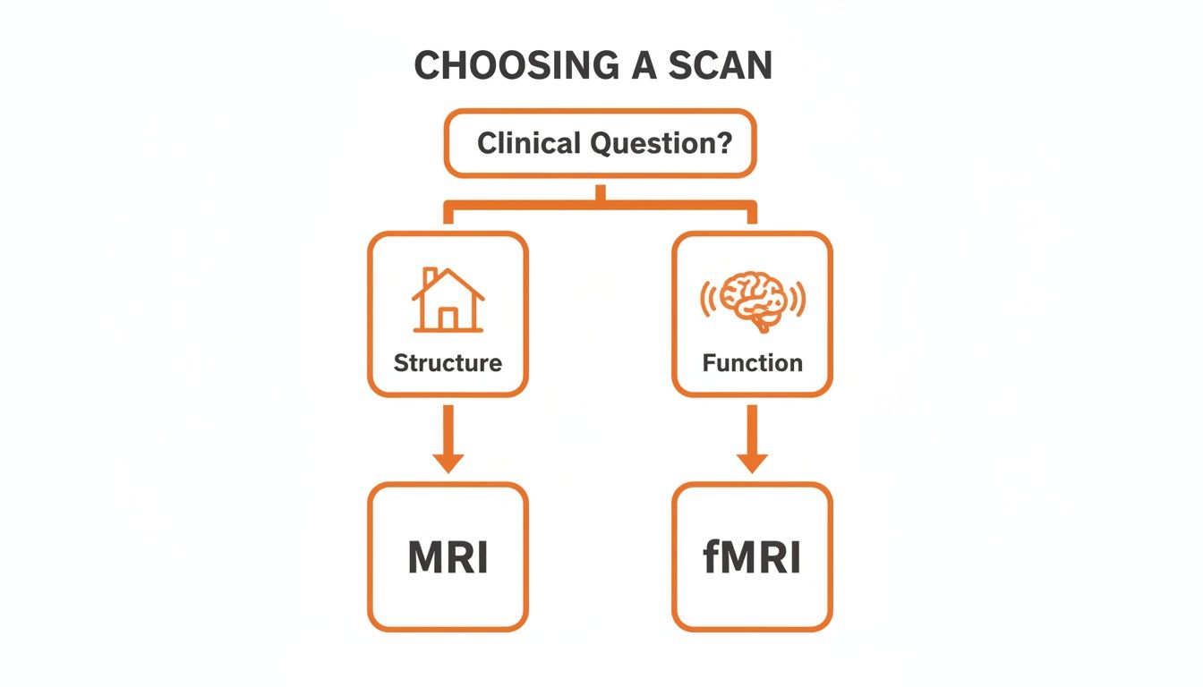
The bottom line is that your clinical question—structure versus function—should always guide your choice of imaging. For post-injury evaluations, this distinction is especially important, as we explore in our guide on complete concussion management.
We're also seeing fMRI move from a pure research instrument to a clinical workhorse. The global fMRI market is expected to reach $1.25 billion by 2031, a testament to its growing role in neurology and psychiatry for pre-surgical planning and evaluating complex cognitive conditions. You can dive deeper into this trend by exploring the latest fMRI market reports.
Weighing the Strengths and Limitations of MRI and fMRI
When you're deciding between an fMRI and an MRI, it's crucial to know that neither one is a magic bullet. Each tool has its own distinct advantages and, just as importantly, significant limitations that you need to consider based on the clinical question you’re trying to answer. The first step is setting realistic expectations about what each scan can—and can't—reveal.
The standout strength of a structural MRI is its incredible spatial resolution. It gives you breathtakingly detailed anatomical pictures, making it the undisputed gold standard for spotting physical issues like tumours, lesions, or the tell-tale signs of a stroke. The big catch? It’s completely static. It provides a perfect "blueprint" of the brain but tells you absolutely nothing about how it’s functioning.
On the other hand, an fMRI’s unique power lies in its ability to map brain activity over time without any invasive procedures. It opens a window into brain function, showing us which areas are firing up during certain tasks. For things like pre-surgical planning or neuroscience research, this functional insight is a complete game-changer.
The Trade-Offs with Functional Imaging
But getting that functional data means making some serious trade-offs. An fMRI’s spatial resolution is much lower than a standard MRI, so the anatomical images are a lot fuzzier. The technique is also incredibly sensitive to a patient's movement. Even tiny shifts can completely corrupt the data, which makes it a real challenge to use with children or patients who are feeling anxious.
Maybe the single biggest hurdle, though, is how complex fMRI data analysis is. The BOLD signal, which is our indirect clue to neural activity, is incredibly faint. Pulling a meaningful signal out of all the background noise requires some heavy-duty statistical processing. This complexity not only leaves more room for error but also demands specialized expertise to interpret the results correctly.
An MRI tells you what is physically there; an fMRI suggests what that structure is doing. This nuanced view is essential for integrating imaging results into a complete clinical picture, preventing over-interpretation of functional data.
The Statistical Demands of fMRI
The statistical heavy lifting required for fMRI is on another level compared to the structural simplicity of an MRI. For instance, research from California's academic institutions has tried to quantify just how much data is needed to get reliable results. To achieve 80% detection power for brain activations, you might only need about 12 subjects for a basic analysis. But that number doubles to 24 subjects once you apply the strict corrections needed for multiple comparisons. You can explore these findings on fMRI statistical power in more detail.
This really shines a light on fMRI's huge data demands—a critical point to remember for both research design and clinical interpretation. Getting a handle on these strengths and weaknesses is the first real step toward using these powerful tools effectively.
To get the most out of any neuroimaging, it's smart to pair it with other objective data. Rapid cognitive assessments can give you a functional baseline, helping you decide if imaging is even necessary and providing crucial context for the results. Discover how Orange Neurosciences can help you build this comprehensive clinical picture by visiting our website or reaching out to our team for more information.
Pairing Neuroimaging with Smart Cognitive Assessments
Neuroimaging is fantastic at showing us the “where” of brain health. It can pinpoint a structural problem or map out which parts of the brain are firing. But on its own, it often misses the “what” and “how well” of a person's cognitive performance. When you combine the anatomical data from an MRI or the functional data from an fMRI with a practical, data-driven cognitive assessment, you get a much clearer, more complete clinical story.
Let’s think about a common scenario: a patient comes in complaining of persistent memory issues and brain fog. Before jumping to a costly and time-consuming fMRI to hunt for functional deficits, a quick, objective cognitive assessment can give you a critical baseline.
This is where a smart assessment tool proves its worth. For example, using a platform like Orange Neurosciences, a clinician can get a detailed cognitive profile in less than 30 minutes. The results might show a specific, measurable weakness in working memory, even if other cognitive abilities seem perfectly fine.
From Broad Search to Targeted Investigation
This kind of objective data does more than just back up a patient's complaints. It gives you the solid information you need to justify ordering advanced imaging and helps you ask the right questions. Instead of going on a broad fishing expedition, the fMRI becomes a focused investigation into the neural networks that support working memory.
This two-step approach has some major advantages:
Justifies Imaging: Having objective cognitive data makes it much easier to make the case for an expensive scan like an fMRI. It helps with insurance approvals and makes sure that these powerful resources are used when they’re truly needed.
Guides Scan Protocol: When radiologists know the exact deficit you’re investigating, they can tailor the fMRI task to specifically test that cognitive function. This boosts the chances of getting truly relevant data.
Provides Context for Results: When you get the imaging results back, you can interpret them alongside the initial cognitive assessment. This directly connects the brain activity patterns you see on the scan to the real-world performance issues the patient is experiencing.
This integrated approach changes neuroimaging from a standalone test into one piece of a much larger diagnostic puzzle. It closes the gap between seeing what the brain is doing and understanding how well it's performing, which is the key to creating treatment plans that actually work.
Combining cognitive measurement with neuroimaging ultimately leads to a more accurate diagnosis and more personalized care. It helps clinicians move past simply managing symptoms and start addressing the root causes of cognitive challenges. By seeing the whole picture, you can build far more effective care plans. You can find out more about how to integrate these tools by exploring our guide on what is cognitive assessment.
Beyond the technical physics, the practical side of ordering an MRI versus an fMRI is where the decision-making rubber really hits the road for clinicians and their patients. Things like cost, who has access to which machine, and what the patient actually goes through during the scan often play a massive role.
Navigating Costs, Availability, and the Patient Experience
Think of a standard structural MRI as a fairly routine procedure. It's widely available in hospitals and imaging centres across Canada, and for medically necessary reasons, it's typically covered by provincial health plans.
Functional MRI, on the other hand, is a different beast entirely. It’s far more expensive and much less common. You’ll usually only find fMRI capabilities in major academic medical centres or highly specialized research hospitals. This scarcity can mean long wait times and significant travel for patients, making it a less practical choice for many routine clinical questions.
The Patient Journey: MRI vs. fMRI
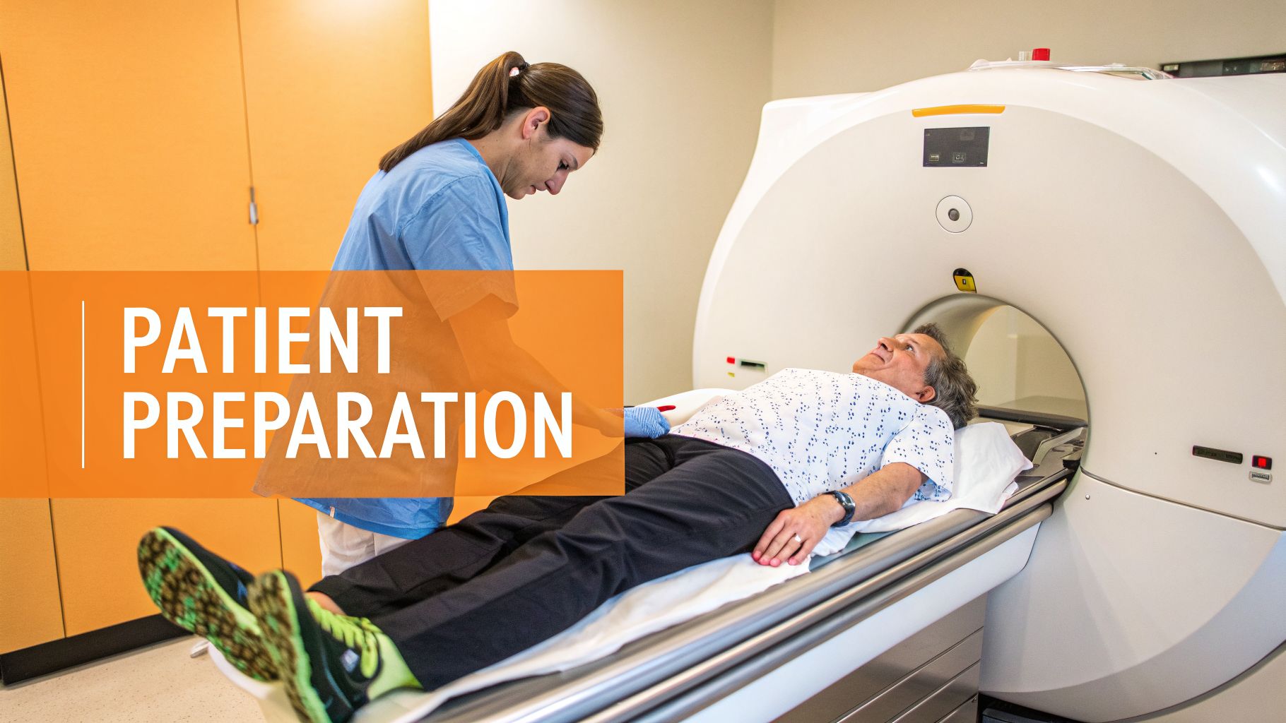
The experience inside the scanner also differs dramatically between an MRI and an fMRI. This is a critical piece of information when preparing someone for imaging. While both scans require the patient to lie still inside the scanner's tube, an fMRI asks for a lot more.
Standard MRI: The main job for the patient is to stay still. That's it. A typical scan runs about 20–45 minutes, and the primary goal is to remain as motionless as possible to get clear pictures.
fMRI Scan: An fMRI session is often a bit longer, anywhere from 20–55 minutes. The key difference is that the patient can't just lie there; they have to actively engage in specific cognitive tasks, like word games or finger-tapping sequences, which are usually displayed on a small screen inside the scanner.
This need for active participation can be a real hurdle for some. Children, anyone with attention challenges, or patients who feel anxious in confined spaces may find it incredibly difficult to follow instructions while also holding perfectly still. Any movement or failure to complete the tasks can unfortunately compromise the quality of the data.
For patients who might struggle with the scanner's environment or the demands of an fMRI, proper preparation is everything. Mock scanner sessions, which recreate the experience without any magnetic fields, can do wonders for a patient's comfort and cooperation, ultimately leading to much higher-quality images.
Taking the time to walk patients through these differences is essential. It helps manage their expectations and ensures the entire process, from the initial referral to the final report, goes more smoothly. A well-prepared patient is far more likely to yield the clear, high-quality data we need for an accurate interpretation, which is a cornerstone of providing genuine client-centred care.
A Few Common Questions About Brain Scans
When it comes to brain imaging, the alphabet soup of MRI and fMRI can be confusing. Let's clear up some of the most common questions we hear from clinicians and their patients.
Which Is Better: MRI or fMRI?
This is a classic "apples and oranges" comparison. Neither scan is better than the other; they just answer entirely different questions.
Think of a structural MRI as the detailed architectural blueprint of the brain. It’s the go-to choice for seeing the physical structure with incredible clarity. If the clinical question is about a suspected tumour, a bleed, or a physical injury, the structural MRI is your tool.
An fMRI, on the other hand, is like watching the electrical grid of a city in real time. It doesn't show the buildings, but it shows which areas are lit up and using power. It tracks blood flow to measure brain activity, making it the superior choice when your question is about function.
The right choice between fMRI versus MRI always comes down to what you need to know: are you looking at the brain's anatomy or its activity?
Can an fMRI Actually Diagnose a Mental Illness?
Not on its own, no. An fMRI isn't a standalone diagnostic test for conditions like depression, anxiety, or ADHD, at least not in a routine clinical setting today. It's a powerhouse for research, helping us understand the neural patterns behind these conditions, but it's not a simple "yes/no" test for diagnosis.
That said, fMRI findings can offer powerful supporting evidence. For example, if a patient with depression shows specific patterns of hypoactivity in the prefrontal cortex, this functional data can add a crucial layer to the diagnostic picture and help fine-tune a treatment plan.
It’s best to think of it as one important piece of a much larger puzzle, not the single answer you’re looking for.
What Is Resting-State fMRI?
Resting-state fMRI (rs-fMRI) is a fascinating technique where we look at the brain's baseline activity. The patient simply lies still in the scanner, not performing any specific task, letting their mind wander.
The goal is to map the brain's functional connectivity—to see how different brain networks are communicating with each other when the brain is in its default "idle" mode. This is incredibly valuable for studying disorders where the core issue might be disrupted communication between brain regions, giving us a window into the brain's fundamental operating system.
Before ordering costly and time-consuming imaging, it’s critical to have objective data on cognitive function. Orange Neurosciences provides rapid, evidence-based cognitive assessments that establish a clear performance baseline. This helps you determine if imaging is the right next step and provides essential context for interpreting any results you get.
Explore how our tools can strengthen your diagnostic process.

Orange Neurosciences' Cognitive Skills Assessments (CSA) are intended as an aid for assessing the cognitive well-being of an individual. In a clinical setting, the CSA results (when interpreted by a qualified healthcare provider) may be used as an aid in determining whether further cognitive evaluation is needed. Orange Neurosciences' brain training programs are designed to promote and encourage overall cognitive health. Orange Neurosciences does not offer any medical diagnosis or treatment of any medical disease or condition. Orange Neurosciences products may also be used for research purposes for any range of cognition-related assessments. If used for research purposes, all use of the product must comply with the appropriate human subjects' procedures as they exist within the researcher's institution and will be the researcher's responsibility. All such human subject protections shall be under the provisions of all applicable sections of the Code of Federal Regulations.


© 2026 by Orange Neurosciences Corporation